
How to Draw Lungs Really Easy Drawing Tutorial
The lungs, which is the organ for respiration is a paired cone shaped organs lying in the thoracic cavity separated from each other by the heart and other structures in the mediastinum. Image: Lungs in situ - anterior view Each. The alveolar walls consist of simple squamous epithelium. Also, the tissues surrounding the alveoli contain.

structure of lungs in hd
lungs How Does Air Enter the Respiratory System? Air enters the respiratory system through the nose or the mouth.

Human Lungs Diagram stock vector art 481669095 iStock
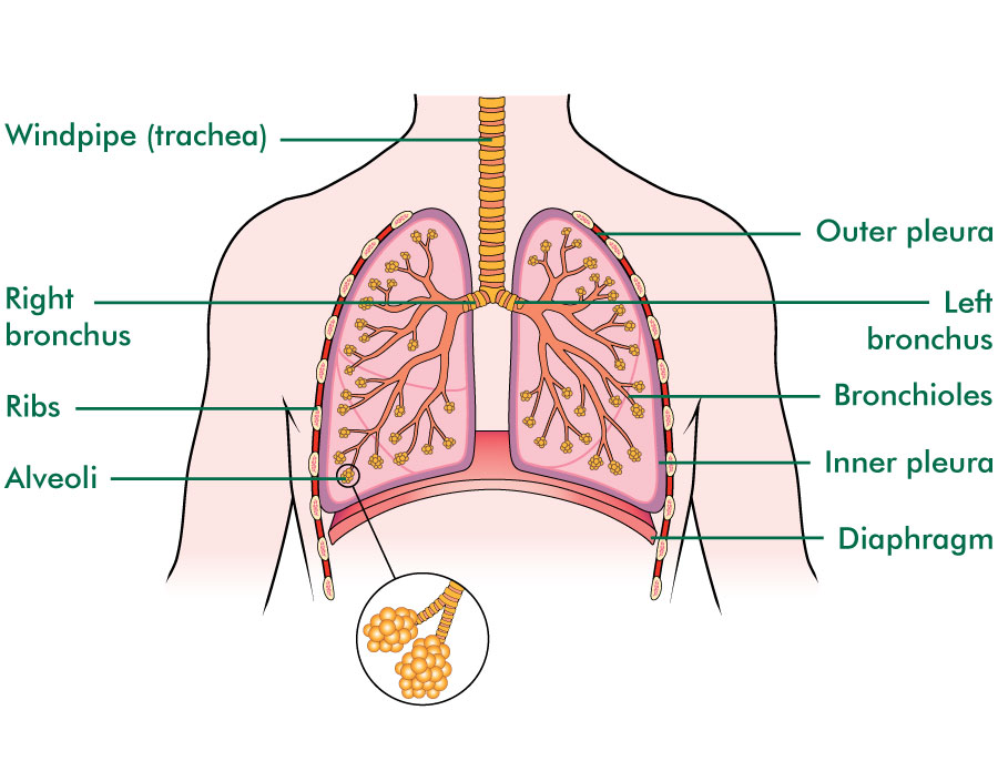
The diaphragm is the flat, dome-shaped muscle located at the base of the lungs and thoracic cavity. The lungs are enclosed by the pleurae, which are attached to the mediastinum. The right lung is shorter and wider than the left lung, and the left lung occupies a smaller volume than the right.

Lung Diagrams Diagram Link
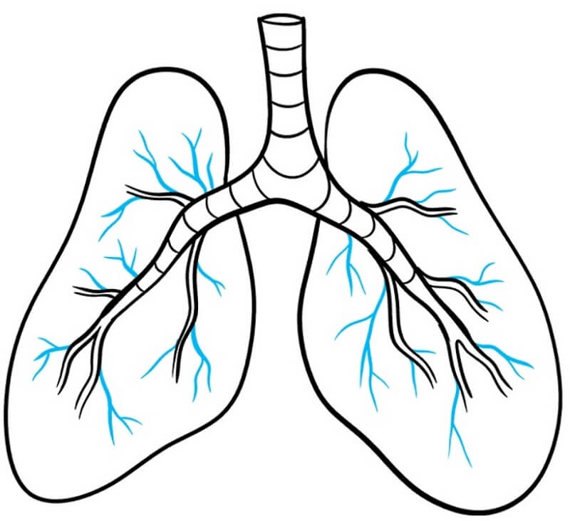
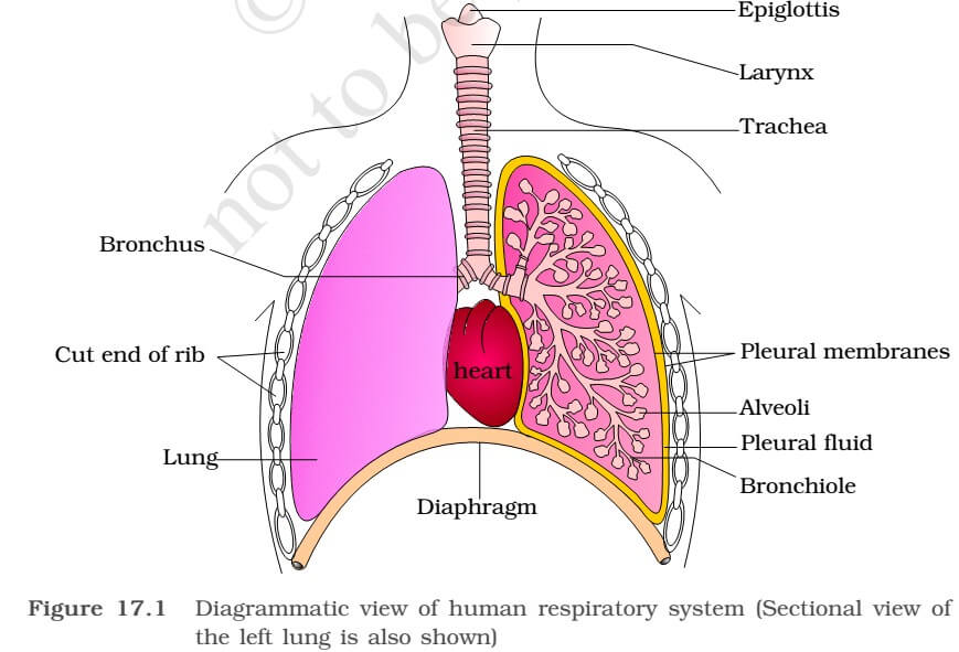
Inside your lungs, the bronchial tubes branch into thousands of thinner tubes called bronchioles. The bronchioles end in clusters of tiny air sacs called alveoli. Air fills your lung's air sacs Your lungs have about 150 million alveoli. Normally, your alveoli are elastic, meaning that their size and shape can change easily.
Lungs PNG Images Transparent Free Download PNGMart
Overview A step-by-step explanation of how your lungs work. What are your lungs? Your lungs make up a large part of your respiratory system, which is the network of organs and tissues that allow you to breathe. You have two lungs, one on each side of your chest, which is also called the thorax.
A Guide to Understand Lung with Diagrams EdrawMax Online
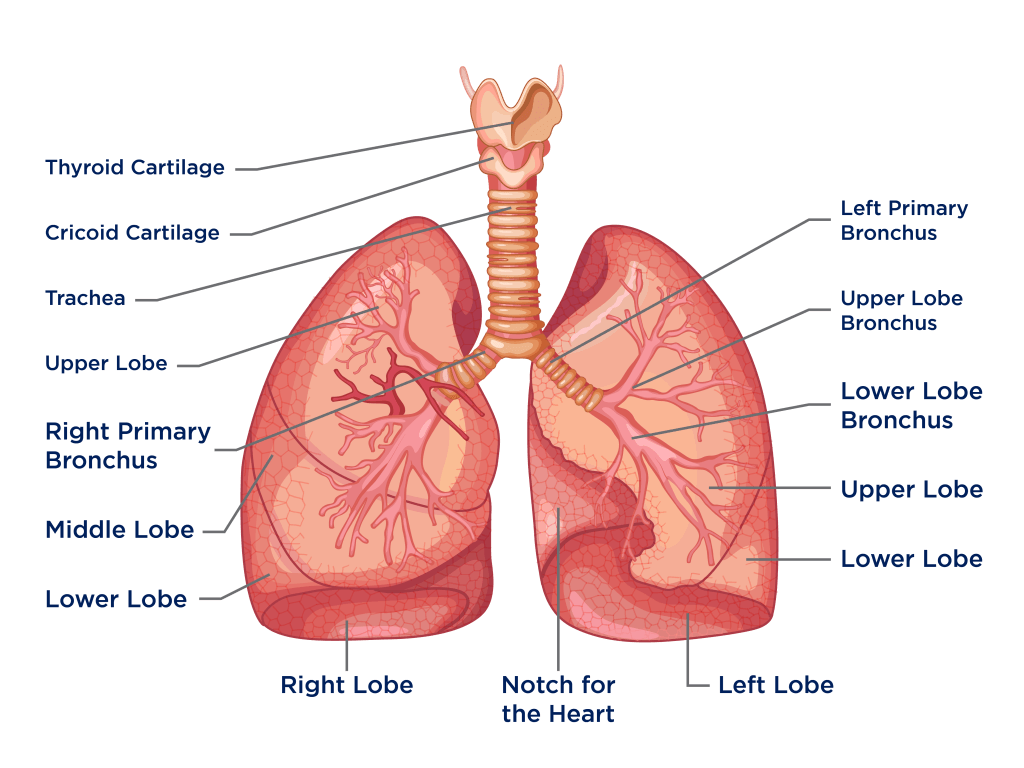
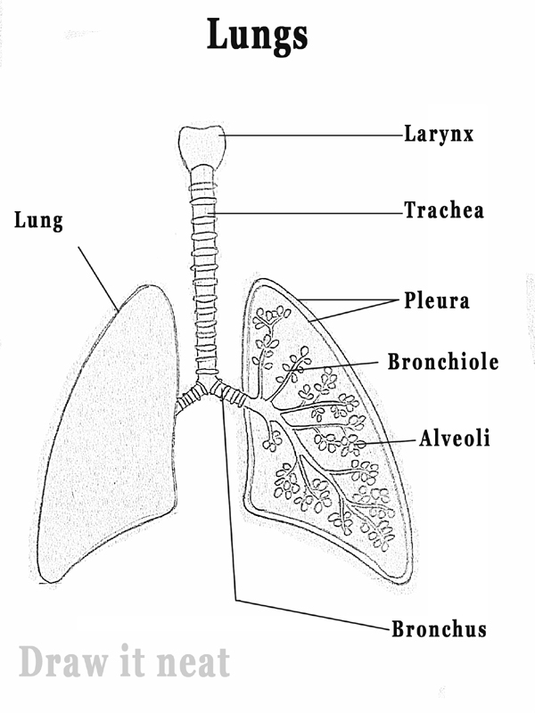
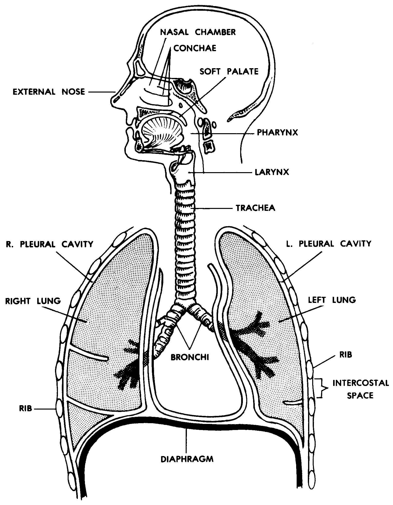
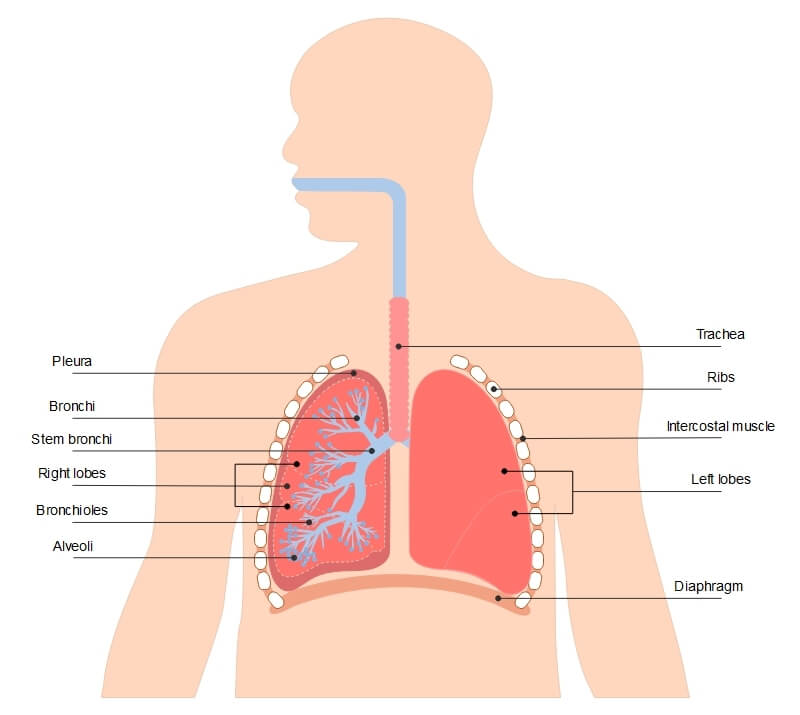
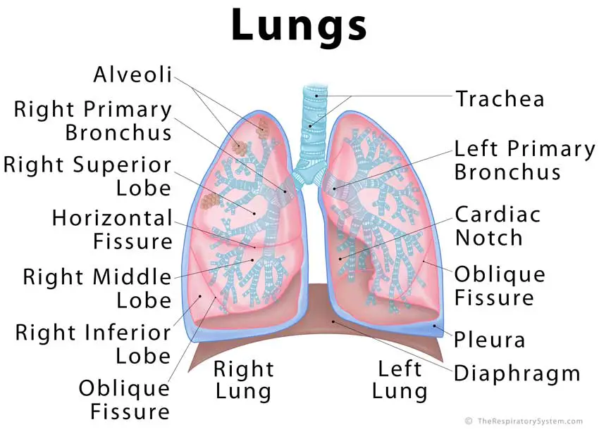
Diagram of the respiratory system. Air enters the body via the nose (preferably) or the mouth. The air enters the main windpipe, called the trachea, and continues en route to each lung via either the right or left bronchus (plural=bronchi). The lungs are separated into sections called lobes, two on the left and three on the right.
How do we breathe? (Lungs and Pleura) Interactive Biology, with Leslie Samuel
Anatomical Position and Relations The lungs lie either side of the mediastinum, within the thoracic cavity. Each lung is surrounded by a pleural cavity, which is formed by the visceral and parietal pleura. They are suspended from the mediastinum by the lung root - a collection of structures entering and leaving the lungs.
DRAW IT NEAT How to draw Lungs diagram
Lung anatomy can get quite complicated extremely quickly. Ease into the topic and cement your knowledge using Kenhub's respiratory system quizzes and labeled diagrams. Regulation of breathing. The breathing cycle is controlled by the respiratory centre located inside the medulla oblongata and the pons of the brain stem. Three major collections.
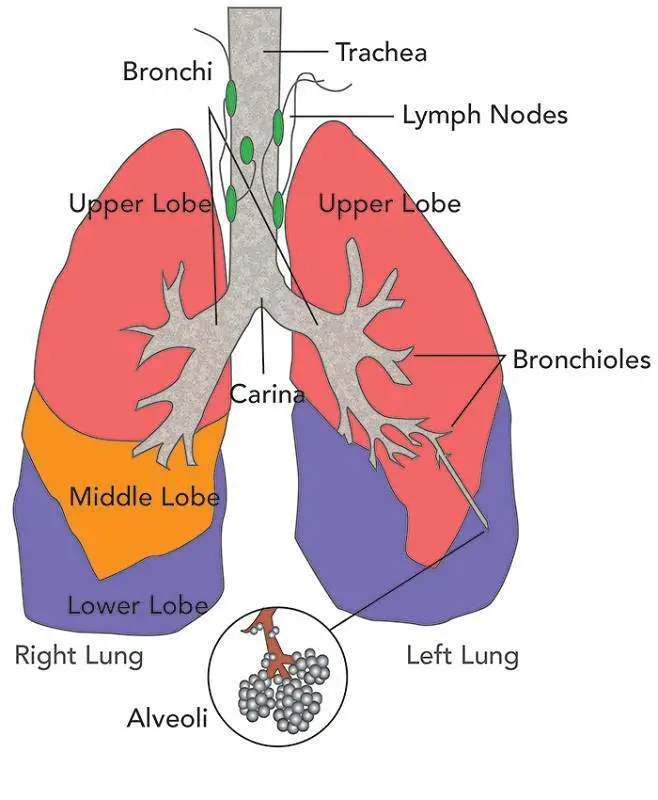
Diagram of lungs
Anatomy Structure There are two lungs (a right and left) in the body, but they are different sizes. The right lung is bigger and is divided into three lobes (separated by fissures), while the left lobe is smaller consisting of two lobes. The left lobe is also smaller as it has to make room for the heart.
Images 07. Respiratory System and Breathing Basic Human Anatomy
Breathing is the term given to the process of taking air into and out of the lungs. The process of inhalation and exhalation Two important structures for breathing are the diaphragm and.
Respiratory System NCERT General Science
Respiratory system diagram The respiratory system How we breathe Respiratory conditions Summary The respiratory system allows air to reach the lungs, from which oxygen enters the blood.

DRAWING OF HUMAN LUNGSDrawing and labelling of human lungs. Easy step to draw for students
Health tips Overview The lungs are the center of the respiratory (breathing) system. Every cell of the body needs oxygen to stay alive and healthy. Your body also needs to get rid of carbon.
A Guide to Understand Lung with Diagrams EdrawMax Online
Lungs-simple diagram of lungs and trachea: Date: 23 December 2006: Source: Patrick J. Lynch, medical illustrator: Author: Patrick J. Lynch, medical illustrator: Permission (Reusing this file) Creative Commons Attribution 2.5 License 2006:
A healthy lung has a pinkish appearance, and if you could see it outside the body, it would look
The lungs are a pair of pyramid-shaped organs in the respiratory system that supply oxygen to the body. They are located in the thorax, with one lung on each side of the chest. They are.
The lungs Lung cancer Macmillan Cancer Support
Respiratory system (Systema respiratorum) The respiratory system, also called the pulmonary system, consists of several organs that function as a whole to oxygenate the body through the process of respiration (breathing).This process involves inhaling air and conducting it to the lungs where gas exchange occurs, in which oxygen is extracted from the air, and carbon dioxide expelled from the body.

Simple Lungs Nursing Respiratory Pinterest Lungs
Simple Lungs diagram The lung, the human gas-exchanging structure, resides in the chest (thorax) wherein its intricate tissues are protected by the muscular and bony thoracic cage. Structure of the Lungs The lungs are seen in the intrathoracic space, which is filled by the mediastinum.
